Leg Anatomy Muscles Ligaments And Tendons / Muscle gross anatomy 12 photos of the muscle gross anatomy gross anatomy of cardiac muscle, gross anatomy of skeletal muscle worksheet, gross muscle anatomy test, muscle gross anatomy quiz, muscular system gross anatomy chapter 10, human muscles, gross anatomy of cardiac muscle, gross anatomy of skeletal.
byAdmin-
0
Leg Anatomy Muscles Ligaments And Tendons / Muscle gross anatomy 12 photos of the muscle gross anatomy gross anatomy of cardiac muscle, gross anatomy of skeletal muscle worksheet, gross muscle anatomy test, muscle gross anatomy quiz, muscular system gross anatomy chapter 10, human muscles, gross anatomy of cardiac muscle, gross anatomy of skeletal.. This important tendon in the back of the calf and ankle connects the plantaris, gastrocnemius, and soleus muscles to. Each of these muscles is a discrete organ constructed of skeletal muscle tissue, blood vessels, tendons, and nerves. Tendons connect the knee bones to the leg muscles that move the knee joint. Muscles, either individually or in groups, are supported by fascia. In the hip, the joint capsule is formed by a group of three strong ligaments that connect the femoral head to the acetabulum.
Ligaments are structures that connect two bones together. There are four major ligaments that surround the knee joint. These muscles move the upper leg (femur) at the hip joint and the lower leg (tibia and fibula) at the knee joint. Ligaments and tendons are fibrous bands of connective tissue that attach to bone. The calf muscle, on the back of the lower leg, is actually made up of two muscles:
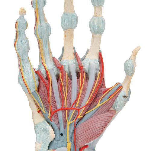
Anatomical Teaching Models Plastic Human Joint Models Hand Skeleton Model With Ligaments And Muscles from www.3bscientific.com These muscles allow the ankle to bend downward and outward. A joint capsule is a watertight sac that surrounds a joint. This lies on the front of the knee and connects the quadriceps muscles of the thigh to the tibia via the patella and patellar ligament (or tendon). Tendons connect the knee bones to the leg muscles that move the knee joint. Ligaments, muscles and tendons keep us connected and help us move. The two main calf muscles, gastrocnemius and soleus, run down the back of the calf and join together to form a strong, thick tendon, the achilles tendon, that attaches to the back of the heel. The foot is a part of vertebrate anatomy which serves the purpose of supporting the animal's weight and allowing for locomotion on land. #muscle and tendon pain in legs #muscles and tendons of the leg and foot #muscles and tendons of the lower leg #muscles ligaments and tendons of the lower leg #muscles tendons and ligaments of the upper leg
These muscles move the upper leg (femur) at the hip joint and the lower leg (tibia and fibula) at the knee joint.
The quadriceps and hamstring muscles work together to straighten (extend) and bend (flex) the leg. #muscle and tendon pain in legs #muscles and tendons of the leg and foot #muscles and tendons of the lower leg #muscles ligaments and tendons of the lower leg #muscles tendons and ligaments of the upper leg The calf muscle, on the back of the lower leg, is actually made up of two muscles: Leg anatomy muscles ligaments and tendons. The calf muscles (gastrocnemius and soleus), which are connected to the calcaneus via the achilles tendon. A muscle strain is a stretch or tear of muscle fibers. The adductor muscles pull the legs together. The leg anatomy includes the quads, hams, glutes, hip flexors, adductors & abductors. Tendons attach muscle to bone. The hamstring muscles in the back of the thigh, the quadriceps muscles in the front, and the adductor (groin) muscles on the inside. To better understand foot and leg muscle/tendon injuries, it is important to appreciate the basic elements that enable your body parts to move. Some of the more common ones are: The gastrocnemius is the larger calf muscle, forming the bulge visible beneath the skin.
Muscles, tendons, and ligaments run along the surfaces of the feet, allowing the complex movements needed for motion and balance. Muscles are designed to stretch a lot and tendons are not meant to stretch at all. There are many muscles located in the lower leg, but there are three that are particularly well known—the gastrocnemius and the soleus, which are the most powerful muscles in the lower leg, and the anterior tibialis. Muscles, either individually or in groups, are supported by fascia. The leg muscles are organized in 3 groups:
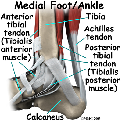
Ankle Anatomy Be In Motion Physiotherapy from www.beinmotion.ca These muscles allow the ankle to bend downward and outward. Muscles, either individually or in groups, are supported by fascia. Flexion, extension, medial rotation, and lateral rotation) and it connects the tibia and the fibula, with the thigh bone (femur). Ligaments are structures that connect two bones together. Leg anatomy muscles ligaments and tendons. The tarsal bones are found near the. There are many muscles located in the lower leg, but there are three that are particularly well known—the gastrocnemius and the soleus, which are the most powerful muscles in the lower leg, and the anterior tibialis. The achilles tendon is also located in the lower leg.
The quadriceps and hamstring muscles work together to straighten (extend) and bend (flex) the leg.
The foot is a part of vertebrate anatomy which serves the purpose of supporting the animal's weight and allowing for locomotion on land. A joint capsule is a watertight sac that surrounds a joint. Major muscles of the ankle. The achilles tendon is also located in the lower leg. The hamstring muscles in the back of the thigh, the quadriceps muscles in the front, and the adductor (groin) muscles on the inside. Posterior view of leg showing muscles and tendons involved in ankle movement. Ligaments and tendons are fibrous bands of connective tissue that attach to bone. The last of the muscle compartments of the lower leg is the lateral compartment (figure 15) is comprised of two muscles, the peroneus longus and the peroneus brevis. The calf muscle, on the back of the lower leg, is actually made up of two muscles: The popliteofibular ligament attaches the popliteus tendon to the fibular head and has a thickness similar to the lateral collateral ligament (fig. Muscle and tendon pain in legs, muscles and tendons of the leg and foot. The adductor muscles pull the legs together. Tendons connect the knee bones to the leg muscles that move the knee joint.
Both cross the ankle, but the peroneus longus wraps underneath the cuboid crossing the plantar aspect of the foot as well, and inserts at the base of the first metatarsal. These muscles allow the ankle to bend downward and outward. The achilles tendon is also located in the lower leg. To better understand foot and leg muscle/tendon injuries, it is important to appreciate the basic elements that enable your body parts to move. Possibly the most important tendon in terms of mobility is the achilles tendon.
Anatomy Of Knee from ix-cdn.b2e5.com To better understand foot and leg muscle/tendon injuries, it is important to appreciate the basic elements that enable your body parts to move. Muscle gross anatomy 12 photos of the muscle gross anatomy gross anatomy of cardiac muscle, gross anatomy of skeletal muscle worksheet, gross muscle anatomy test, muscle gross anatomy quiz, muscular system gross anatomy chapter 10, human muscles, gross anatomy of cardiac muscle, gross anatomy of skeletal. Leg anatomy muscles ligaments and tendons. Muscles, either individually or in groups, are supported by fascia. Muscles, tendons, and ligaments run along the surfaces of the feet, allowing the complex movements needed for motion and balance. The peroneal muscles (peroneus longus and peroneus brevis), on the outside edge of the ankle and foot. This important tendon in the back of the calf and ankle connects the plantaris, gastrocnemius, and soleus muscles to. To ensure your body moves smoothly with a minimum of friction, muscles are enveloped in a slippery skin like tissue called fascia.
Ligaments are soft tissue structures that connect bones to bones.
The calf muscles (gastrocnemius and soleus), which are connected to the calcaneus via the achilles tendon. Some of the more common ones are: The peroneal muscles (peroneus longus and peroneus brevis), on the outside edge of the ankle and foot. In the leg, muscle strains happen when a muscle is either stretched beyond its limits or forced into extreme contraction. Ligaments are soft tissue structures that connect bones to bones. Two of these ligaments are in the center of the joint, and they cross each other. Muscle and tendon pain in legs, muscles and tendons of the leg and foot. The soft tissue in the knee joint (tendons, ligaments, menisci, cartilage) that provides stability in the knee and hold the bones. In the hip, the joint capsule is formed by a group of three strong ligaments that connect the femoral head to the acetabulum. These muscles allow the ankle to bend downward and outward. These are called the cruciate ligaments and consist of the anterior cruciate ligament and the posterior cruciate ligament. The two main calf muscles, gastrocnemius and soleus, run down the back of the calf and join together to form a strong, thick tendon, the achilles tendon, that attaches to the back of the heel. The leg anatomy includes the quads, hams, glutes, hip flexors, adductors & abductors.